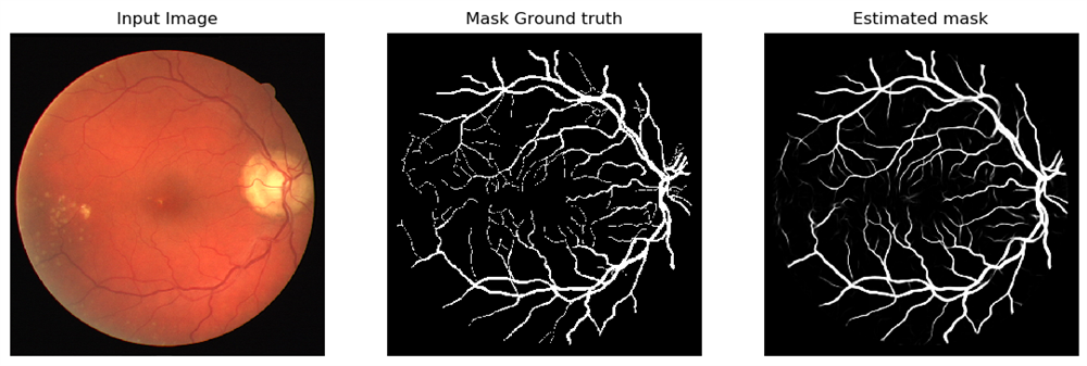
Medical image segmentation is an important research topic in the field of medical image analysis. It utilizes computer vision and machine learning techniques to automatically or semi-automatically segment target structures in medical images, providing support for disease diagnosis, treatment, and monitoring. Currently, deep learning-based methods such as convolutional neural networks (CNNs), recurrent neural networks (RNNs), and attention mechanisms have achieved significant success in medical image segmentation. Traditional methods based on image features, morphology, and statistics, as well as emerging technologies such as multi-modal information integration, uncertainty learning, and augmented reality, have also been applied in medical image segmentation.
Our research focuses on optimizing deep learning-based segmentation frameworks and their applications to clinical practice. Firstly, fine segmentation of human organ structures can assist doctors in disease diagnosis. For example, segmentation of structures such as blood vessels, lungs, and hearts can help doctors determine the morphology, location, and size of lesions, guiding clinical decision-making. Secondly, we also aim to integrate segmentation results with surgical navigation systems, allowing doctors to accurately locate target structures during surgery, improving surgical safety and precision.

Related Projects
- Diagnosis and surgical planning for pediatric congenital heart disease: In collaboration with Shanghai Children's Medical Center, this project has two main tasks. The first task is to diagnose pediatric congenital heart diseases, including Tetralogy of Fallot, single ventricle, and anomalous pulmonary venous drainage. The second task is to plan for Fontan surgery, a surgical repair for single ventricle heart defects, and assess postoperative risks.
Selected Papers
 |
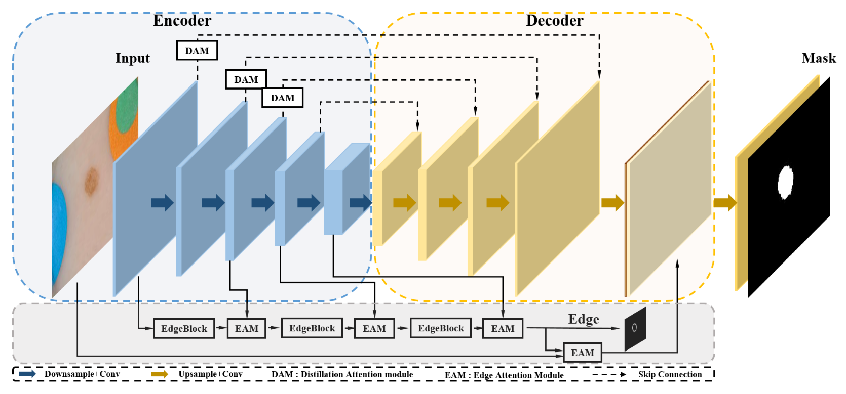
AEC-Net: Attention and Edge Constraint Network for Medical Image Segmentation EMBC 2020 [BibTex] |